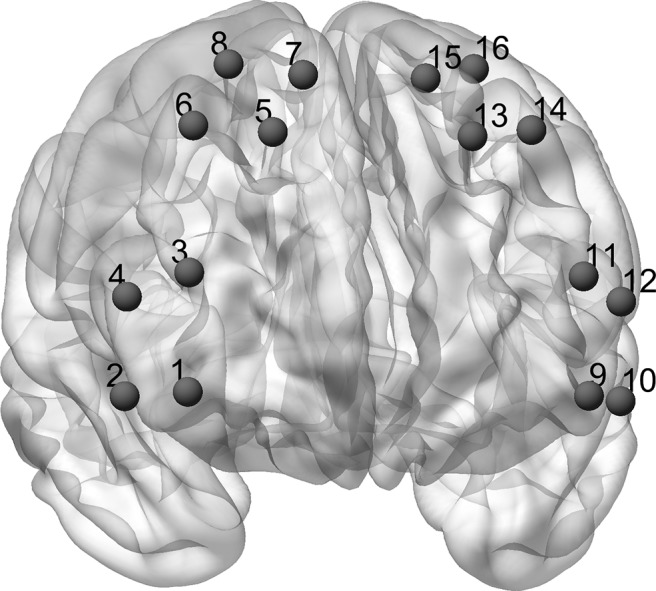
Figure 2.

fNIRS channel positions. The channel setup covered parts of the lateral and medial PFC. For final analysis, we averaged over all channels. The Matlab toolbox NFRI (Singh et al., 2005) was used to estimate the Montreal Neurologic Institute coordinates corresponding to the international EEG 10–20 positions. Channel positions were visualized using BrainNet Viewer (Xia et al., 2013).
