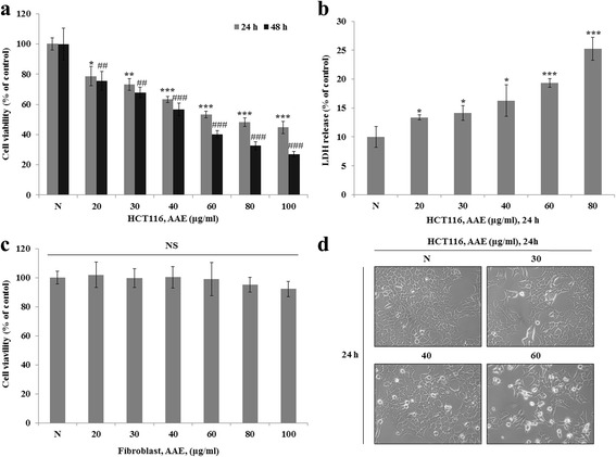
Fig. 1.

AAE reduces cell proliferation in HCT116 colon cancer cells. a, c Cell viability was measured by MTT assay. a is HCT116 cell line. c is Fibroblast cell line. The statistical analysis of the data was carried out by use of an T-test. */#p < 0.05, **/##'p < 0.01 and ***/###p < 0.001 (each experiment, n = 3). b LDH assay was performed for assessing cell deaths. Cytotoxicity was induced by AAE. The statistical analysis of the data was carried out by use of an T-test. *p < 0.05, **p < 0.01 and ***p < 0.001 (each experiment, n = 3). d AAE affects the morphology of HCT116 cells, and promotes cell death in a dose-dependent manner. HCT116 were treated with AAE (0, 30, 40, and 60 ìg/ml) for 24 h
