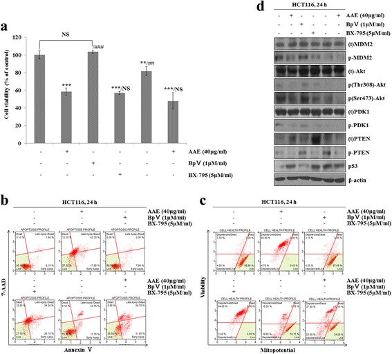
Fig. 7.

AAE induces apoptosis by regulating the phosphorylation of PDK1 and Akt through the PTEN/p53-independent pathway. Cells were treated with 1 μM BpV, 5 μM BX-795 and 40 μg/ml AAE for 24 h. a Cells viability was measured by MTT assay. The statistical analysis of the data was carried out by use of an an T-test. */#p < 0.05, **/##p < 0.01 and ***/###p < 0.001 (each experiment, n = 3). b Apoptotic effects of different concentration AAE were evaluated by Muse™ Annexin V and Dead Cell Assay Kit. HCT116 were treated with 1 μM BpV, 5 μM BX-795 and 40 μg/ml AAE for 24 h. c Mitochondria membrane potential were evaluated by Muse™ Mitopotential kit. HCT116 were treated with 1 μM BpV, 5 μM BX-795 and 40 μg/ml AAE for 24 h. for 24 h. d Cells were treated with 1 μM BpV, 5 μM BX-795 and 40 μg/ml AAE for 24 h. for 6 h. The expression of PTEN, PDK1, Akt (Thr308 and Ser473), MDM2, p53 were analyzed by western blot analysis
