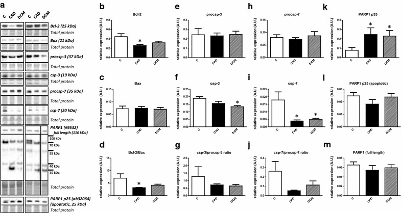
Fig. 2.

Lack of significant changes in the expression of apoptotic markers in human end-stage heart failure. a Representative immunoblots of Bcl-2, Bax, procsp-3, csp-3 and PARP1 in control (C) and heart failure samples due to myocardial infarction (CAD) and dilated cardiomyopathy (DCM). The right PARP1 blot and its corresponding total protein stain section are spliced because of marker lane interference. b, c, e, f, h–m Quantification of Bcl-2, Bax, procsp-3, csp-3, procsp-7, csp-7, PARP1 p35, PARP1 p25 and total PARP1 immunoblots. d, g Quantification of Bcl-2/Bax, csp-3/procsp-3 and csp-7/procsp-7 ratios. Data are presented as mean ± SEM. *P < 0.05 vs. C. n = 4, 6, 10 for C, CAD and DCM respectively
