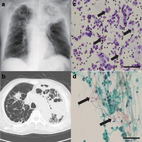
Fig. 1.

a, b Chest X-ray (a) and computed tomography (b) on POD 25 showed massive consolidation in the left upper lobe and a nodular shadow in the right upper lobe. c Eosinophilic infiltration (arrow) was confirmed by BAL fluid cytology using Diff-Quik stain. d Filamentous fungi were observed in the BAL fluid cytology with Papanicolaou staining
