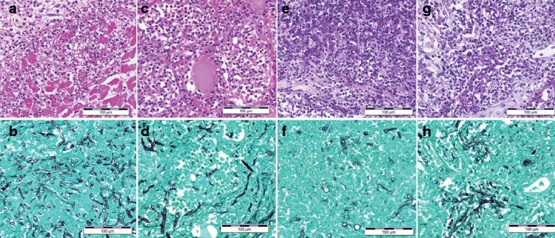
Fig. 3.

Histological findings in the (a, b) heart, (c, d) thyroid gland, (e, f) spleen, and (g, h) kidney. HE staining (a, c, e, g) and Grocott staining (b, d, f, h) are shown (bar = 100 μm). Filamentous fungi with the infiltration of inflammatory cells were also observed in these organs
