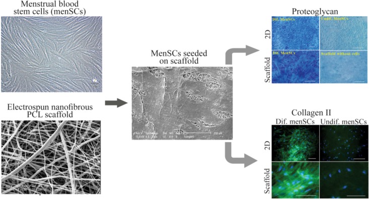
Figure 2.
Culture and chondrogenic differentiation of MenSCs on nanofibrous scaffold. The image analyses of the scanning electron microscopy show that cells penetrated and adhered well on the surface of the mesh. Development of cartilage-like tissue in cultured constructs has been examined histologically with respect to the presence of proteoglycan and collagen type II (Scale bar: 100 μm). PCL: Polycaprolactone, Dif: Differentiated, 2D: Two Dimensional. (Adopted from Kazemnejad et al 2014 40, with minor modification).

