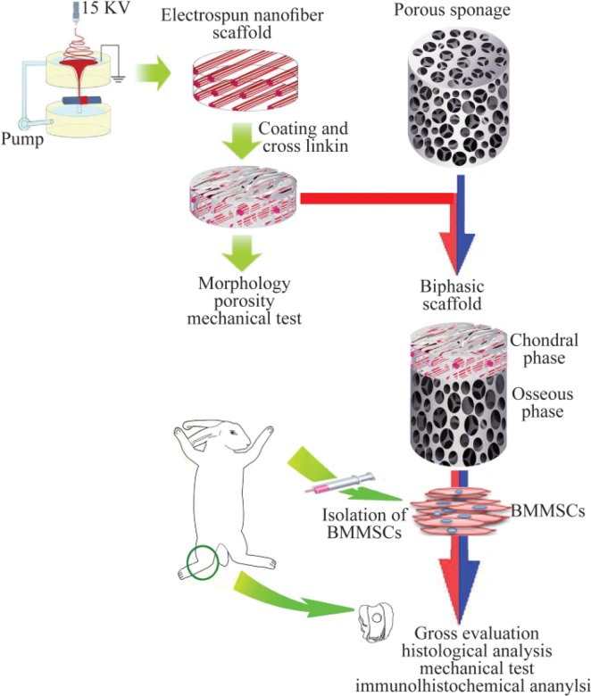
Figure 3.
A schematic model for in vivo study on repair of osteochondral defects using constructs composed of nanofibers and stem cells. The nanofiber is considered as the chondral phase. Porous sponage is used as the osseous phase. After combining with BMMSCs, biphasic complex was utilized to repair osteochondral defects in the animal model (Adopted from Liu et al 201456, with modification).

