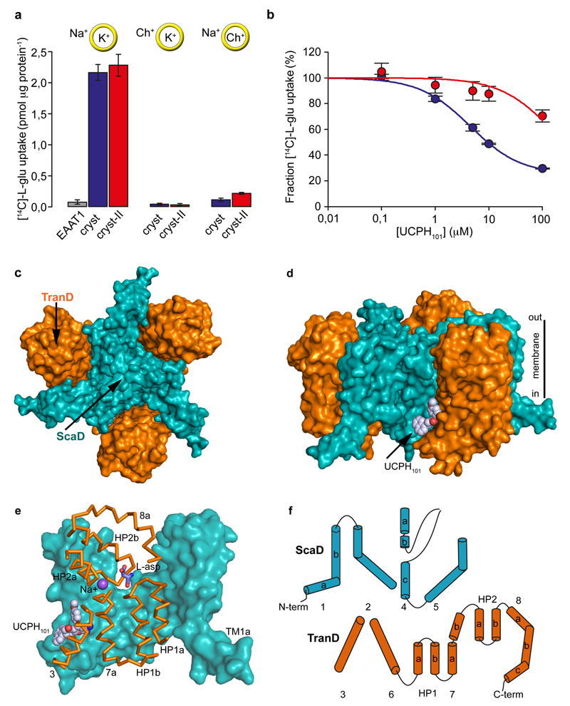
Figure 1. Function and architecture of EAAT1cryst.
a-b, Uptake of radioactive L-glutamate by purified EAAT1 (grey), EAAT1cryst (blue), and EAAT1cryst-II (red) reconstituted in liposomes. Transport was abolished when choline (Ch+) was used in the extra- or intra-liposomal solutions (yellow circles) (a). UCPH101 inhibits glutamate transport in a concentration dependent manner (b). Plots depict an average of three independent experiments performed with duplicate measurements, and error bars represent s.e.m. c-d, Structure of EAAT1cryst trimer viewed from the extracellular solution (c) and from the membrane (d) highlighting the ScaD (teal) and TranD (orange). e, EAAT1cryst monomer viewed parallel to the membrane. The ScaD domain is represented as surface (teal), and several helices and loops in the TranD (orange) have been removed. f, Domain organization diagram of EAAT1cryst monomer.

