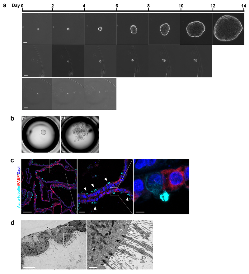
Figure 5. Human endometrial organoids have clonogenic ability and are bipotent.
(a) Phase-contrast images of (from top to bottom row): an organoid forming from a single cell; a single cell forming a spheroid with no further growth, and a single cell showing no growth. Images were taken every two days. Scale bar, 50 μm. Experiment was performed with 3 clonal lines derived from 2 endometrial and 1 decidual organoid cultures.
(b) Representative image showing expansion of a clonal culture at passage 1 (p1) from a single organoid (at passage 0, p0) in a 96-well. Scale bar, 500 μm. 12 clonal cultures were established from organoids from 5 different patient samples (4 endometrial-derived and 1 decidual-derived).
(c) IF on clonally-derived endometrial organoid cultures subjected to the full cocktail of hormonal stimuli to visualize two main endometrial epithelial cell types: ciliated cells (acetylated α-tubulin) (cyan) and secretory cells (PAEP) (red). Scale bars from left to right: 100 μm, 20 μm and 5 μm. Representative of 4 clonal lines derived from 2 different endometrial organoid cultures.
(d) EM on clonally-derived endometrial organoid cultures subjected to the full cocktail of hormonal stimuli showing basal bodies of fully formed cilia. Scale bars: 10 μm and 1 μm. Experiment performed twice using 1 clonal endometrial organoid culture.

