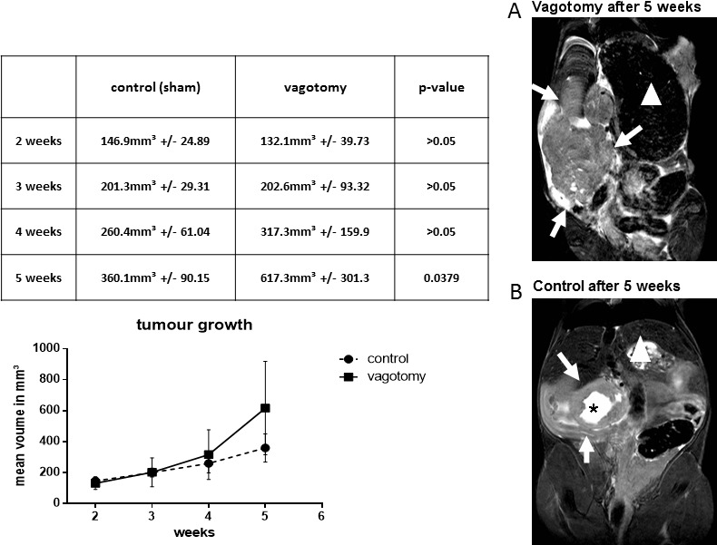
Figure 1. Vagotomy led to significantly increased tumor growth.

Tumor bearing mice were scanned in a high field 7.0 Tesla MRI scanner for small animals. Vagotomy led to an increased tumor volume 5 weeks after tumor implantation (control (360.1 ± 90.15 mm3, n = 8) versus vagotomy (617.3 ± 301.3 mm3, n = 7, p < 0.05). MRI images (high resolution T2-TSE images of the coronal plane) of the vagotomy group show a more irregular disseminated pattern (A), whereas MR images of mice with sham-operation resembled a more spherical growth pattern (B), often showing a central zone of necrosis (black asterisk). White arrows indicate the tumor. In addition, the large stomach after vagotomy is readily identifiable (stomachs are marked with a white triangle).
