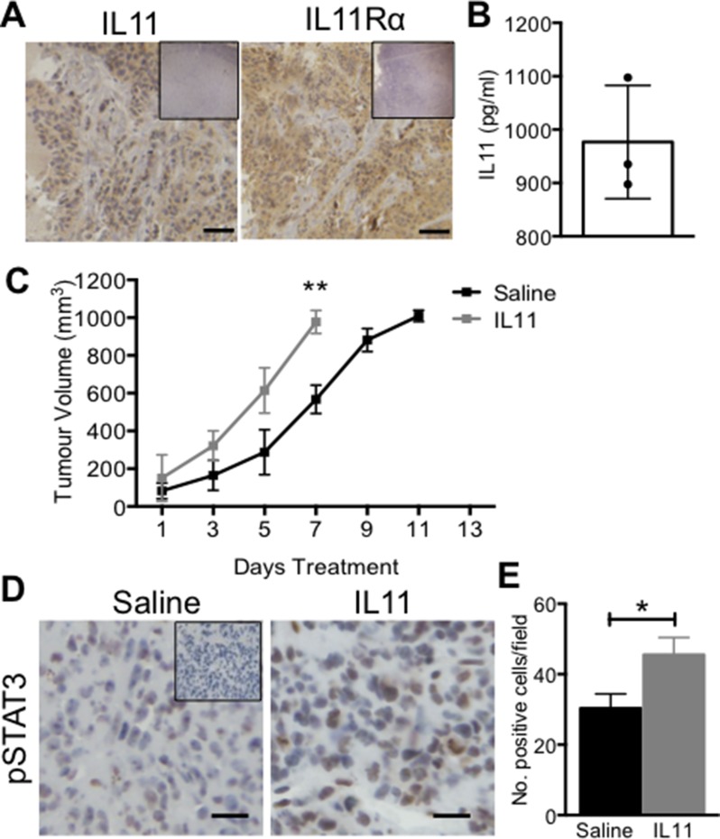
Figure 3. The effect of IL11 on AN3CA xenograft tumour growth in vivo.
Female Balb/c athymic nude mice were inoculated with 5 × 10⁵ AN3CA cells on both hind flanks. (A) Immunohistochemistry for IL11 and IL11Rα was performed on untreated AN3CA xenograft tumour tissue sections (n = 3). Brown indicates positive staining with blue nuclear counterstain. Bars represent 50 μm. Insets are negative controls. (B) IL11 protein (pg/ml) was quantified in ANC3A tumours by ELISA. Data are mean ± SEM of triplicate experiments (n = 3). (C) Mice with established AN3CA xenograft tumours were administered with saline vehicle control or IL11 (500 μg/kg) three times weekly and tumour volume calculated. (D) Representative photomicrographs of pSTAT3 immunohistochemistry on AN3CA subcutaneous control or IL11-treated tumour sections. Bars represent 50 μm. Inset is negative control. (E) Total number of pSTAT3 positive cells per field (×20 magnification) were counted from 5 fields per tumour. (D, E) Data are mean ± SEM. t-test; *p < 0.05, **p < 0.01 (n = 3/group).

