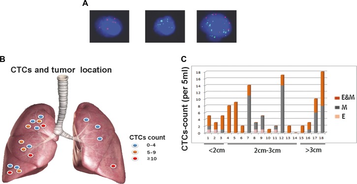
Figure 1. EMT-CTCs prior to operation in cohort A with early stage lung adenocarcinoma.
(A) From left to right, RNA-in situ hybridization clearly identified EMT markers in an E-CTC (red dots), a M-CTC (green dots) and an E&M-CTC(red and green dots), respectively. Therefore, EMT-CTCs were classified as E-CTCs, M-CTCs and E&M-CTCs, respectively. (B) In the scatter diagram, highly abundant total CTCs (E-CTCs, M-CTCs and E&M-CTCs) was prone to being in the tumors in right side, e.g., case A2(CTCs count = 17), case A10(CTCs count = 14) and case A18(CTCs count = 18). (C) Eighteen patients were divided into three groups by tumor size, i.e., < 2 cm, 2cm-3cm and > 3 cm, respectively. The different subtypes of CTCs, i.e., E-, M- and E&M-CTCs were marked by different colors, respectively. M-CTCs were prone to being in the tumors > 2 cm. Furthermore, E-CTCs were prone to being in the tumors < 3 cm.

