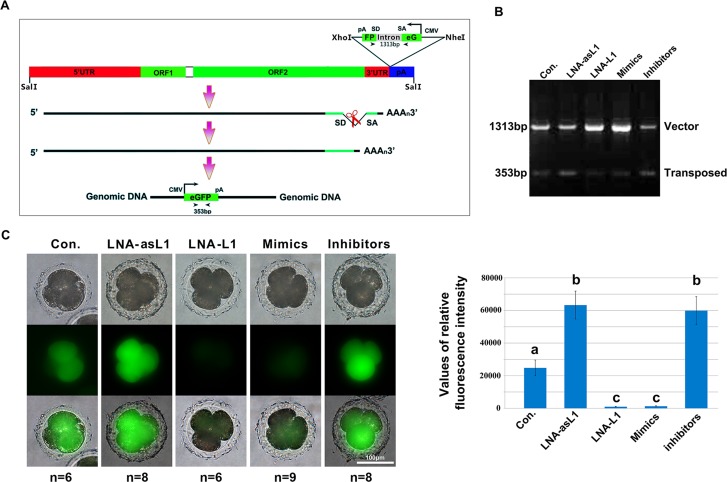
Figure 4. L1-siRNAs control L1 retrotransposition.
(A) Schematic diagrams of the pCEP4-pL1-eGFP expression cassettes used for L1 retrotransposition assays. This L1 retrotransposon contains an intron-interrupted eGFP reporter in the 3′ UTR region with its own CMV promoter and polyadenylation signal. The eGFP indicator cassette is in an antisense orientation relative to L1. Only when eGFP is transcribed from the L1 promoter, spliced, reverse transcribed and integrated into the genome does embryo become eGFP positive. Arrowheads depict the primers used in PCR based genomic DNA analysis. SD, splice donor; SA, splice acceptor; (B) PCR analysis of retrotransposition events in 4-cell stage embryos. The primers, flanking the intron in eGFP, were used for PCR amplification of genomic DNA, and the obtained PCR products of 1313 bp (corresponding to the intron-containing vector) and 353 bp (corresponding to the retrotransposed insertion that lacks the 960 bp intron) are shown; (C) Fluorescence detection of retrotransposition events in 4-cell stage embryos. Error bars represent s.d.. Values with different superscripts differ significantly (p < 0.001).

