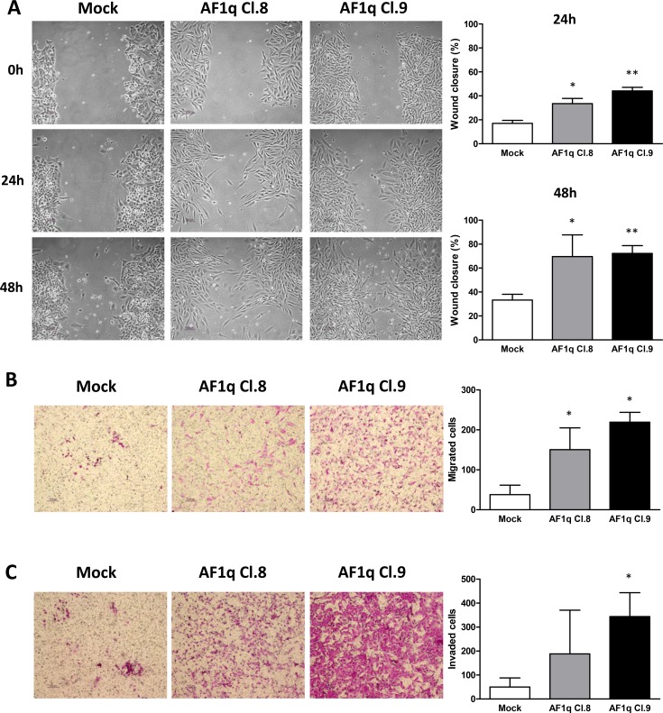
Figure 3. AF1q stable overexpression promotes cell motility and migration in A2780 ovarian cancer cells.
Whound-healing (A), migration (B) and invasion (C) assays evaluating the change of cell mobility in A2780 cells stably overexpressing AF1q (AF1q Cl.8 and 9) compared to mock cells. (A) Representative images of wound healing assays evaluated at 24 and 48 h after scratch. Graphs represent the quantification of “gap closure” (Cl.8: p = 0.013 and 0.049 at 24 and 48 h, respectively; Cl.9: p = 0.006 and 0.002 at 24 and 48 h, respectively). (B) and (C) Representative images and graphs relative to transwell migration (p = 0.027 and 0.015 for Cl.8 and Cl.9, respectively) and invasion (p = 0.024 for Cl.9) assays, evaluated at 48 h, graphs represent the relative migration ability calculated from at least 4 fields under a light microscope. The data are represented as mean ± S.D. from three independent experiments.

