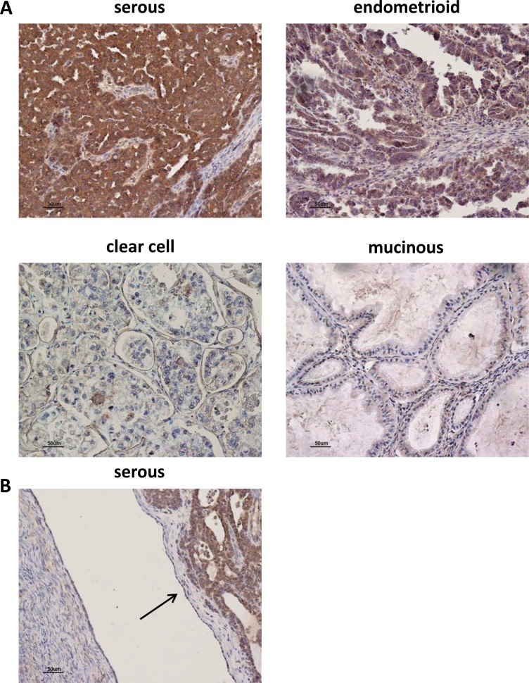
Figure 7. AF1q immunostaining in human ovarian tumor tissues.
(A) Representative images of AF1q staining in human ovarian tumors of different hystotypes. High IHC staining of AF1q in serous and endometrioid tumors. Low and negative IHC staining of AF1q in clear cell, and mucinous tumors, respectively. (B) Representative image of negative IHC staining of AF1q in OSE cells indicated by the arrow.

