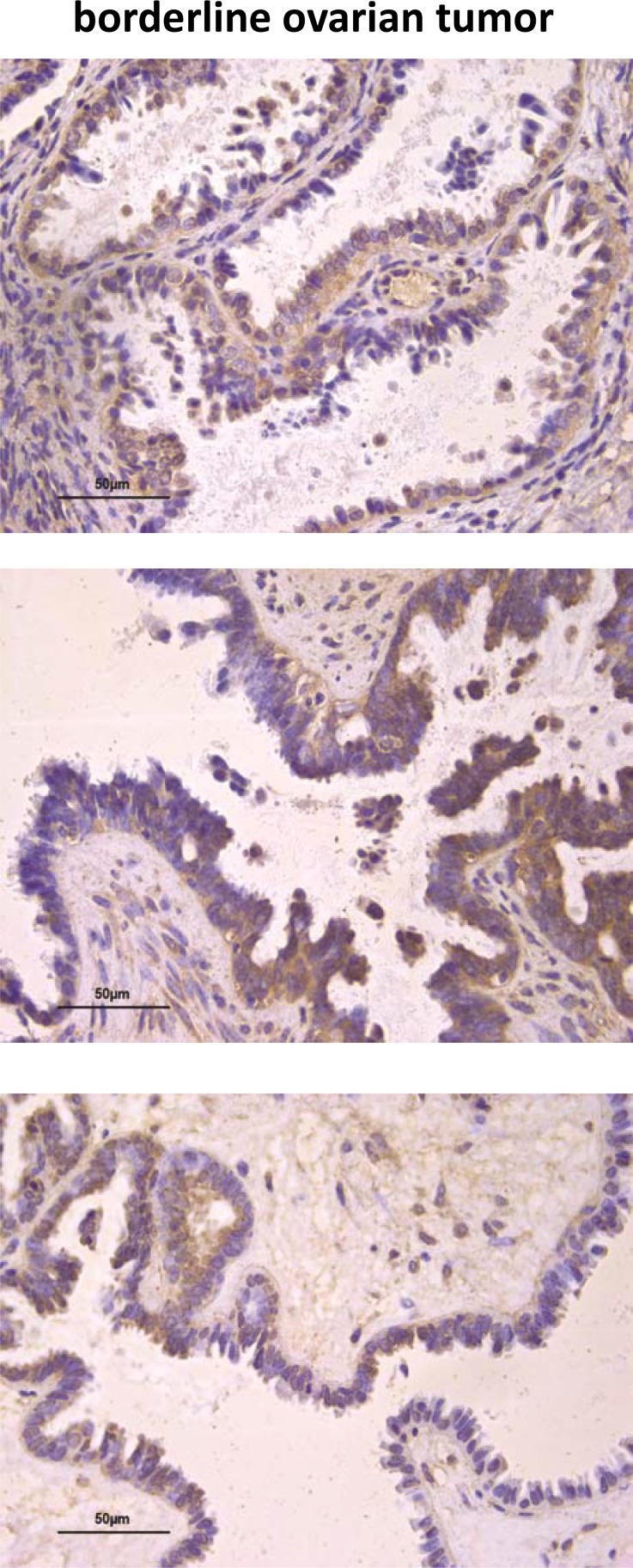
Figure 8. AF1q immunostaining in human ovarian serous BOT.
Examples of IHC staining for AF1q in BOT tumor cells (upper panel). Example of heterogeneous protein staining in BOT cells (middle panel). Negative IHC staining of AF1q in areas of BOT without evidence of atypical epithelium, which becomes detectable in the areas of transition between normal epithelium and atypical epithelial proliferation (lower panel).

