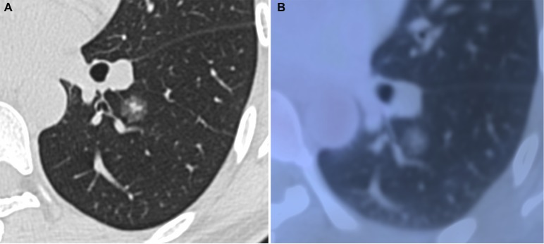
Figure 2. Minimally invasive adenocarcinoma in 40-year-old woman.
(A) transverse lung-window HRCT scan demonstrates a round, spiculated, well-defined parenchymal mixed GGN with pleural indentation in the apical segment of the left lower lobe. (B) PET/CT fusion image with perfusion mode shows a 11.9-mm mixed GGN with 0.25 of CTR and 0.81 of SUVmax.

