
Figure 2. Positive expression of mTOR or beclin1 in the hippocampus of each group 24h after cerebral ischemia (immunohistochemical staining, 400x).
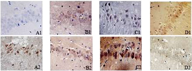
A1–D1: immunohistochemical results of beclin1 in the hippocampus for SO, I/R, IH+I/R and inhibitor group respectively. A2–D2: immunohistochemical results of mTOR in the hippocampus for SO, I/R, IH+I/R and inhibitor group respectively.