
Figure 5. HBx-ΔC1 regulated liver CSC properties through Stat3-Nanog signaling.
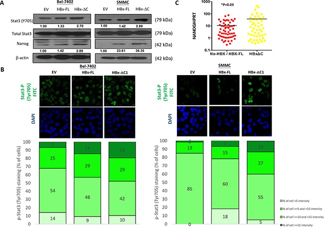
A. Western blot analysis revealed that HBx-ΔC1 expressing Bel-7402 and SMMC-7721 cells had increased Stat3 phosphorylation (Y705) and expression of Nanog, when compared to respective EV and HBx-FL expressing cells. B. Increased fluorescent nuclear staining of p-Stat3 (Y705) was observed in HBx-ΔC1 cells in both Bel-7402 and SMMC-7721when compared with respective EV and HBx-FL, as analyzed by immunefluorescence (IF) staining (40X objective). Stained cells were classified into 4 groups as per the fluorescent intensity measured by Photoshop CS5. C. qPCR analysis demonstrated that the expression of Nanog was up-regulated in HCC samples detected with HBx-ΔC1 (n=48) when compared with those with HBx-negative and HBx-FL (n=59) (*p=0.03, t test).