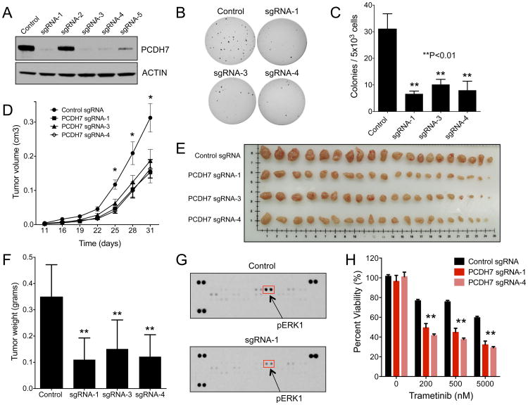
Figure 4. Genetic inactivation of PCDH7 suppresses tumor growth and sensitizes NSCLC cells to MAPK inhibitors.
A, Western blot analysis of PCDH7 in H1944 KRAS mutant NSCLC cells infected with a lentivirus expressing Cas9 and a control sgRNA or sgRNAs targeting PCDH7. B-C, Soft agar colony formation of PCDH7 knockout H1944 cells (B) and associated quantification (C) (**p<0.01 vs. control, one-way ANOVA test). D, Quantification of tumor volume over time (*p<0.05 vs. control, Student's t-test). E, Tumors from NSG mice injected with H1944 cells with control or PCDH7 sgRNA lentiviruses. F, Quantification of tumor weights shown in (E), (**p<0.01 vs. control, Student's t-test). G, Phospho-MAPK antibody array demonstrating that PCDH7 knockout tumors exhibit reduced ERK activation. H, Cell viability assay demonstrating that PCDH7 knockout in KRAS mutant cells enhanced sensitivity to the MEK inhibitor Trametinib (*p<0.05, **p<0.01 vs. control, Student's t-test).

