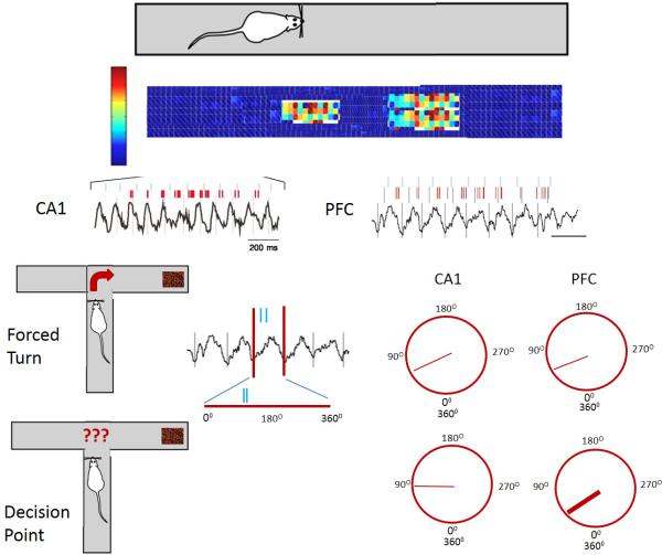
Fig. 4.
Example of hippocampal theta modulation of PFC units. Rat is running on a linear track. Two place cells from the hippocampus are recorded as evidenced by increased firing rates of cells as the animal ran through the place fields. APs from the cells are indicated by vertical blue and red lines positioned above the theta rhythm. In the PFC the APs are temporally linked to the APs in the hippocampus although there is a time delay between the APs in the hippocampus and those in the PFC. The modulation of PFC APs by hippocampal theta occurs even in the absence of PFC theta. In a T-maze animals have to make either a forced turn (top figure) or decided whether to go left or right to receive a food award (cocoa puffs). The theta wave can be drawn as both a straight line as well as a circle to represent the 360O of the theta wave. When the animal has a forced turn there is a preferred phase (straight line within the circle) for the APs to occur in both CA1 and PFC. When the animal has to make a decision there is a much stronger phase lock of PFC neurons with the hippocampal theta as evidenced by a thicker line. Figures are based on work by Siapas et al. (Siapas et al., 2005) and Jones et al. (Jones and Wilson, 2005).

