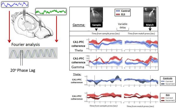
Fig. 5.
Relationship of coherence with cognition. EEG spectral coherence represents the consistency of the phase difference between two EEG signals when compared over time. In a delayed non-match to sample study in rats with a history seizures electrodes were placed in the PFC and hippocampus. Coherences between the waveforms in the hippocampus and PFC were calculated. Coherence is a measure of synchronization or coupling between two EEG signals and is based mainly on the conformity of phase differences between the EEG signals. The two waves shown from the PFC and hippocampus are of the same amplitude and frequency, but there is a phase shift. Phase differences are typically measured in degrees where a complete cycle is 360 degrees. In this example, the phase difference is approximately 30 degrees. Coherences are dynamically measured when controls and rats with early-life seizures (ELS) are doing the test. Data on left is time-linked to the sample press, and data on the right to the match press, revealing functional differences between control (blue) and ELS (red) rats when the envelopes diverge. ELS rats showed increased theta and gamma coherences following the sample press. The lower panel show dynamic CA1-PFC theta coherence in correct and incorrect trials. In trials with 20-30 second delays, control rats did not show performance-related differences in CA1-PFC coherence between correct (light blue) and incorrect (dark blue). In trials with 20-30 second delays, ELS rats showed increased CA1-PFC coherence in correct trials (light red) relative to incorrect trials (dark red) particularly around the time of the sample press and prior to the match press (arrows). Modified from Kleen et al. (Kleen et al., 2011b) with permission.

