
FIGURE 5.
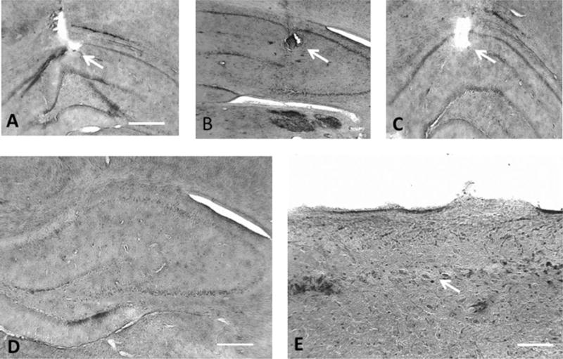
Histology and electrode placement. A–C demonstrates electrode placement in three rats (arrows). The electrode tip was seen in the struatum radiatum/stratum lacunosum moleculare in all animals. D demonstrates wide spread cell loss in one TLE rat. In E a region of CA1 from a TLE shows cell loss (arrow). Photomicrogaphs modified with Abobe Photoshop© to eliminate nonmorphological artifact. Scale = 1 mm (A–D), 400 μm for E.