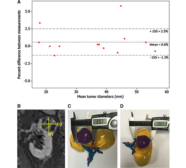
Fig. 3.

3D models accurately depict tumor size. (a) Bland-Altman plot of diameter measurements made on 2D images versus 3D models. The fact that the points lie around the mean demonstrates that there is no inherent bias between the two methods. (b) Coronal MRI showing two diameter measurements (D1 = 39.1mm and D2 =37.5mm). (c) Diameter measurement D1 of 3D printed model by calipers. (d) Diameter measurement D2 measured on the same 3D printed model.
