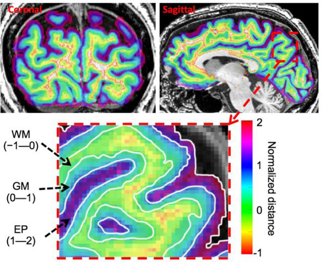
Figure 1.
Example of normalized depth map on coronal and sagittal slices for a subject. Coronal slice locations are in posterior occipital lobe close to the functional slice prescription. Sagittal slices are near the mid-sagittal plane to clearly show the calcarine sulcus. Right: enlargement of sagittal slice showing normalized depth coordinate ranges for WM, GM, and EP compartments.

