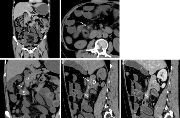
Figure 1.
A computed tomography (CT) scan showing the distended and fluid-filled small bowel (A), which is consistent with small bowel obstruction. An unenhanced axial CT image just below the duodenum shows a tiny high density dot surrounded by soft tissue density (B:arrow). Reconstructed coronal (C) and sagittal (D) images afford a better understanding of the linear high density structure (arrow). Our preoperative diagnosis was an accidentally ingested fish bone penetrating the duodenum and causing small bowel obstruction. Interestingly, the foreign body was obscured on enhanced CT due to the influence of inflammatory and normal tissue enhancement (E).D: duodenum,S: small intestine

