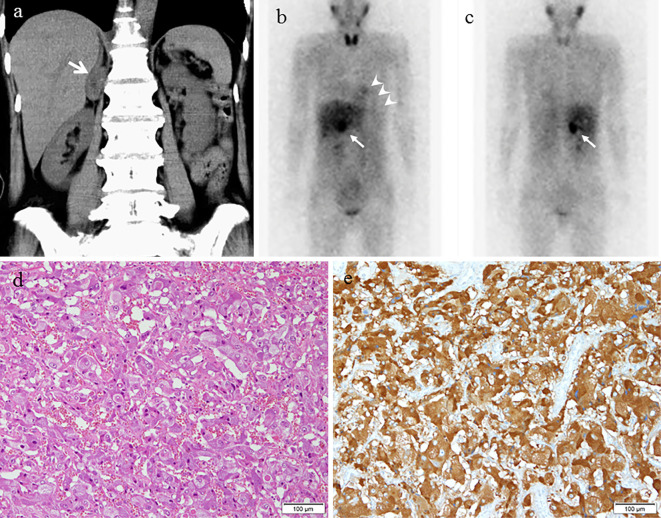
Figure 5.
CT, 123I-MIBG scintigraphy, and the histopathology of the adrenal tumor. A 30-mm right adrenal tumor (arrows) is observed on CT (a). The strong uptake of the adrenal tumor is observed on 123I-MIBG scintigraphy (b: anterior view, c: posterior view) (arrows). uptake of MIBG in the myocardium is low (arrowheads). The histopathological findings are consistent with pheochromocytoma (d: Hematoxylin and Eosin staining, scale bar: 50 μm, e: immunostaining with chromogranin A).

