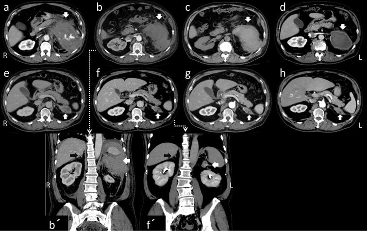
Figure 1.
Abdominal CT scans of the axial lesion, taken on the 1st (a), 3rd (b), 10th (c), 49th (d), 196th (e), 389th (f), 531st (g), and 679th (h) days [on the 3rd (b) and 389th (f) days, with coronal sections (b’) (f’) ]. Except on the 10th day (c), all of the images are enhanced. The left peritoneal mass gradually decreased, and continuity with the left adrenal gland became apparent (white arrows). The right adrenal gland had a normal shape (black arrows in b’, f’).

