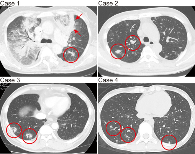
Figure.
Chest computed tomography images of pulmonary mucormycosis-suspected findings in Cases 1-4. The red circles in the images of Cases 1, 2, and 4 show multiple small nodules. The red arrows in the image of Case 1 show diffuse infiltration partly surrounded by a thick, wall-like consolidation. The red circles in the image of Case 3 show two nodules with the reversed halo sign.

