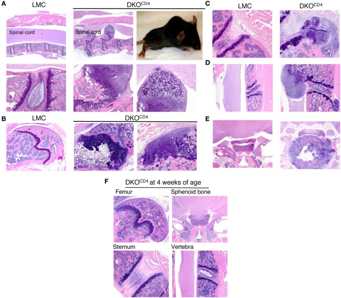
Figure 2.
DKOCD4 mice exhibit ectopic accumulation of chondrocytes. Bones were removed from LMC and 129.DKOCD4 mice, fixed, decalcified, and stained with hematoxylin and eosin (H&E). (A) Sections of spinal column [2× (top) and 10× (bottom)], including a photograph of the paralyzed DKOCD4 mouse from which they were obtained, are shown. Other sections displayed include the (B) femur [4× and 10× (far right)], (C) sternum (10×), (D) vertebra (4×), and (E) sphenoid bone (2×). (F) H&E analysis of bones is depicted from 4-week-old 129.DKOCD4 mice, showing femur (4×), sphenoid bone (2×), sternum (10×), and vertebra (4×). Images are representative of at least five mice.

