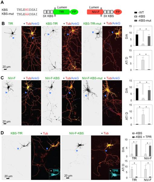
Figure 5. Fusion of a KLC-binding sequence to somatodendritic proteins causes missorting of the chimeras to the axon.
(A) Three copies of the KLC-binding sequence (KBS) from SKIP (Pernigo et al., 2013) or an inactive WD-to-AA mutant of it (KBS-mut) were fused to the cytosolic N-terminus of TfR or C-terminus of NiV-F, both tagged with fluorescent proteins (FP) (GFP or mCherry). (B, C) DIV7 neurons co-expressing mCherry-tubulin (Tub) (red) with TfR-GFP (B) or NiV-F-GFP (C), WT (left) or fused to KBS (middle) or KBS-mut (right) were immunostained for AnkG (blue). Enlarged regions of the somatodendritic domain and axon tips are shown in Figure S3. Dendrite/axon (D/A) and axon tip/dendrite (AT/D) ratios were quantified from 25 neurons and expressed as mean ± SD. * P<0.001. (D) DIV7 neurons co-expressing KBS-TfR-GFP (left) or NiV-F-KBS-GFP (right), mCherry-tubulin (Tub) (red) and the dominant-negative mutant KLC TPR-HA (cyan). Dendrite/axon (D/A) and axon tip/dendrite (AT/D) ratios were quantified from 25 neurons and expressed as mean ± SD. * P<0.001. In all images, arrowheads point to the AIS.

