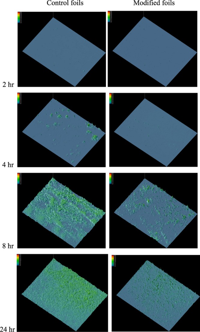
Figure 5.

Inhibition of colony formation on the DAP-Cys-TEG-NPrSi-O-Ti6Al4V surfaces. The foils were incubated at 37 °C. Colonization of bacteria on the surface of Ti6Al4V foils was recorded after 2, 4, 8, and 24 h incubation. Following incubation, the foils were washed seven times with PBS, then stained for 20 min at rt with a live/dead BacLight kit to visualize live bacteria by green fluorescence, with nonmodified Ti6Al4V foils as the controls. Three independent sets of foils were analyzed. Representative fluorescence surface plots of colonies, processed to appear as 3D colorized peaks, are shown at 40× magnification.
