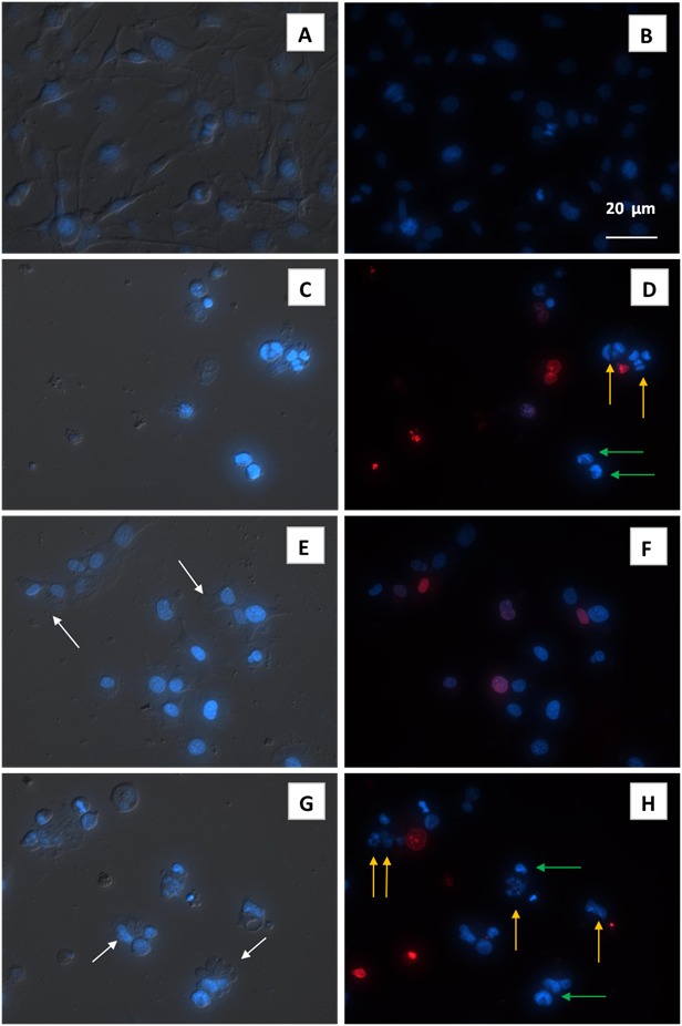
Figure 4.

Cytomorphological characteristics (fluorescence microscopy) of human glioma U251 cells treated for 48 h with 4-thiazolidinone derivative Les-3288 (E, F), doxorubicine (Dox) (C, D), and temozolomide (TMZ) (G, H); control – (A, B). Left – DIC image of treated cells. Les-3288 and Dox were used in 1 ug/mL dose, while TMZ was used in 10 µg/mL dose. Right – fluorescent image of treated cells (blue color – staining with fluorescent DNA-specific dye Hoechst-33342, red color – staining of damaged cells with the ethydium bromide. White arrows - plasma membrane blebbing, green arrows - condensed chromatin, yellow arrows - nucleus fragmentation.
