
Figure 5.
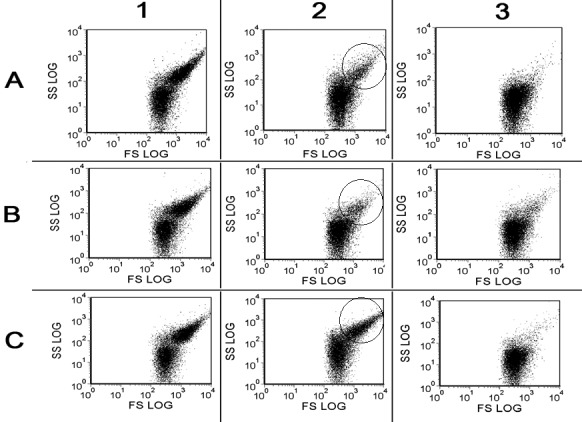
Flow cytometry of washed human platelets activated by 0.125 NIH/mL of thrombin. 1-3 – 1, 2, and 3 minutes after the stimulation, respectively. A. – control probe; B. – in the presence of 50 μg/mL of platelet aggregation inhibitor from the Echis multisquamatis (PAIEM) snake venom; C. – in the presence of 1% dimethyl sulfoxide as the inhibitor of platelet activation. SS – side light scattering, parameter of platelets granulation: FS – frontal light scattering, parameter of platelets shape. Traces are typical for 3 independent experiments made in triplicate.