

Abstract
Background:
This is to assess one of the rare complications after total knee replacement and to assess risk factors of failure.
Methods:
11 patients with varus knee and an average age of 67 years underwent TKA between 2005 and 2013. All patients returned with a sudden sharp knee pain, disability to walk and significant decrease in ROM about 4 to 8 weeks after surgery. Radiographic examination revealed a lateral femoral condylar stress fracture.
Results:
After analyzing the images, we found common characteristics among all patients, which might be attributable to the later fracture including varus deformity>25, femoral component lateralization, and valgus correction.
Conclusion:
Surgeons should be aware of the risk factors to consider before, during, and after surgery.
Keywords: Lateral femoral condylar fracture, Osteoporosis, Severe varus deformity, Stress fracture, Total knee arthroplasty
Introduction
Various complications of total knee arthroplasty (TKA) including infection, periprosthetic fractures, stress fractures, implant failure, loosening of implants, and dislocation are becoming more widely recognized as the number of reported patients and the length of follow-up increases (1-3). Stress fracture of the distal femur – fractures above the femoral component – is an uncommon but one of the challenging complications that may happen following a TKA (4). Fractures of the femur in the presence of a total knee arthroplasty may occur intraoperatively or postoperatively. Management of these fractures is often challenging because of a variety of factors, including those related to the fracture itself, and the quality of the bone, prosthesis, and patient. Most postoperative fractures result from acute trauma, but stress fractures without any trauma are also encountered (1-7).
Stress fractures represent a fatigue failure of bone, occurring with a spectrum of severity of structural injury, and healing potential varies by location (8). There is no comprehensive classification system for stress fractures incorporating both clinical and radiographic characteristics of the injury that is applicable to all bones. Stress fracture can begin as an increased number or size of micro cracks. These micro cracks can coalesce or propagate to create a frank fracture line (1-10).
The incidence of stress fracture is increasing due to several factors including longer life expectancy, greater activity levels, and higher rate of revision arthroplasty,. What is important is understanding that stress fracture occur in regions of stress concentration adjacent to the femoral component and that the presence of the prosthesis has a significant effect on fracture treatment (1-14).
Patients and Methods
We reviewed the clinical and radiographic records of 11 patients including 9 women and 2 men with an average age of 67 years after sustaining a lateral femoral condyle stress fracture following TKA between 2005 and 2013. All of them had radiographic signs of osteopenia of lateral femoral condyle with varus deformity of > 25 degrees before surgery. Mean preoperative IKDC score was 43 (range, 32 to 53), which was improved to 75 (range, 69 to 81) at the latest follow up. Eight patients were overweight (BMI>35). Knee alignment was neuter in 8 patients while 2-4 degrees of valgus alignment was achieved in 3 patients. All patients returned with a sudden sharp knee pain, inability to walk, and significant decrease in rage of motion 4-8 weeks after the index surgery. Radiographic examination revealed a lateral femoral condyle stress fracture. We observed 3 types of fracture patterns. Type I had mild valgus alignment with no obvious lateral condyle displacement (case I), type II had obvious lateral condyle displacement with severe valgus alignment (case II), and type III had a bicondylar fracture (case III).
All patients had been candidate for revision TKA using constrained prostheses (NexGen Legacy Constrained Condylar Knee, Zimmer Inc., Warsaw, Indiana). Postoperative follow-up ranged from 27 to 39 months (mean, 32 months).
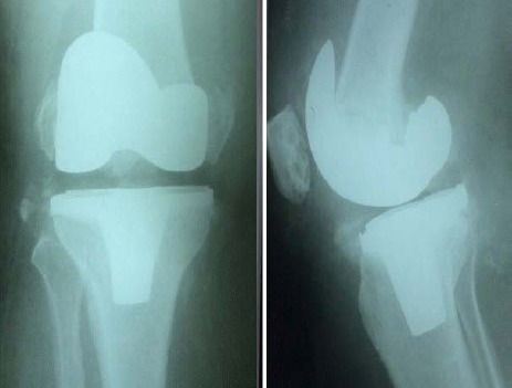
Case 1
A 68-year-old woman, BMI=23, with 27° of varus deformity in the right knee and thrust on weight bearing underwent primary TKA [Figure 1]. Patient returned after 4 weeks with a sharp knee pain and inability to walk. Clinical examination of the collateral ligaments was unremarkable but severe lateral femoral condylar fracture was observed in radiographs [Figure 2]. The patient underwent a revision arthroplasty using a stemmed femoral component retaining the tibial component polyethylene [Figure 3].
Figure 1.

Post operative radiographs after a primary TKA.
Figure 2.

Post operative radiographs 4 weeks after primary TKA. Condylar fracture is notable on the lateral side of the femur.
Figure 3.

Postoperative radiographs after revision arthroplasty.
Case 2
A 73-year-old woman, BMI=35, varus deformity of 30°, and stiffness in the left knee underwent primary TKA. She returned with lateral femoral condyle fracture after 8 weeks, and underwent a revision arthroplasty using. stemmed femoral and tibial components, constrained polyethylene and TM metaphysial femoral cone [Figures 4; 5].
Figure 4.

Post operative radiographs 8 weeks after primary TKA. Lateral femoral condylar stress fracture is notable.
Figure 5.

Post operative radiographs after revision arthroplasty.
Case 3
A 60-year-old woman, BMI=39, varus deformity of 25° with thrust on weight bearing underwent primary TKA [Figure 6]. She returned with lateral femoral condyle fracture after 8 weeks, and underwent a revision arthroplasty using. stemmed femoral and tibial components, constrained polyethylene and TM metaphysial femoral cone [Figures 7; 8].
Figure 6.

A 60-year-old female with 25 degrees of of left knee varus deformity and thrust.
Figure 7.
Lateral and anteroposterior views showing bicondylar femoral stress fracture 8 weeks after primary TKA.
Figure 8.

Post operative radiographs after revision arthroplasty using a constrained prosthesis and metaphysial cone.
Discussion
In our patients, we found common characteristics including varus deformity of greater than 25 degrees, gait with varus thrust, BMI>35, excessive lateralization of the femoral component, excessive alignment correction to reach normal to mild valgus alignment, and localized osteopenia of lateral femoral condyle in the pre-operative radiographs.
Varus deformity along with lateral thrust unloads the lateral compartment of the knee resulting in osteopenia of the lateral femoral condyle over the years. By correction or over-correction of the deformity, loads are transferred to the osteopenic lateral femoral condyle immediately after surgery. Reynolds noted that alignment in the coronal plane is an important biomechanical factor in the etiology and healing of these stress fractures (15). Osteopenia results in lower fatigue strength of the bone and stiffness of the adjacent bone due to arthritis, which are undoubtedly important etiologic factors (16, 17).
Improvements in quality of life and life expectancy have resulted in substantial increase in the rate of total knee arthroplasty (TKA) and thus the likelihood of postoperative complications. Orthopedic surgeons should be aware of possible challenging risks of lateral femoral condyle stress fracture after TKA (18). Pre-operative awareness of the risk factors might be preventive over the rehabilitation period. For this reason, we recommend embedding femoral prosthesis as medial as possible while avoiding patellar maltracking, utilizing femoral stem extension for additional fixation, controlled weight bearing after surgery, retaining 3 to 5 degrees of varus alignment, and impaction of various types of bone grafts in the lateral femoral condyle. Our data is limited to few numbers of patients that does not allow drawing a definite conclusion. However, future studies can be designed in the light of current findings.
References
- 1.Ayers DC, Dennis DA, Johanson NA, Pellegrini VD. Instructional course lectures, the american academy of orthopaedic surgeons-common complications of total knee arthroplasty. J Bone Joint Surg Am. 1997;79(2):278-311. Ayers DC, Dennis DA, Johanson NA, et al. Common complications of total knee arthroplasty. J Bone Joint Surg Am 1997; 79 278-311. [Google Scholar]
- 2.Jiann- Long Jean L, Chian-Her, Lee CH, Ru-Yu Pan RY, Chang JH, Chern TC et al. stress Stress fracture of the proximal tibia after total knee arthroplasty: a case report. J Formos Med Assoc. 2001;100(8):561–564. [PubMed] [Google Scholar]
- 3.Engh GA, Ammeen DJ. Periprosthetic. fractures adjacent to total knee implants: treatment and clinical results . Instr Course Lect. 1998;47:437–48. J Bone Joint Surg Am 1997; 79: 1100-1113. [PubMed] [Google Scholar]
- 4.Phil McGraw P, Arun Kumar A. Periprosthetic fractures of the femur after total knee arthroplasty. J Orthop Traumatol. 2010;11(3):135–141. doi: 10.1007/s10195-010-0099-6. [DOI] [PMC free article] [PubMed] [Google Scholar]
- 5.Whitehouse MR, Mehendale S. Periprosthetic fractures around the knee: current concepts and advances in management. Curr Rev Musculoskelet Med. Curr Rev Musculoskelet Med. 2014;7(2):136–144. doi: 10.1007/s12178-014-9216-0. [DOI] [PMC free article] [PubMed] [Google Scholar]
- 6.Platzer P1, Schuster R, Aldrian S, Prosquill S, Krumboeck A, Zehetgruber I, et al. Management and outcome of periprosthetic fractures after total knee arthroplasty. J Trauma. J Trauma. 2010 Jun;68(6):1464–1470. doi: 10.1097/TA.0b013e3181d53f81. [DOI] [PubMed] [Google Scholar]
- 7.Jae DooYoo JD, Nam Ki Kim NK. Periprosthetic fractures following total knee arthroplasty. Knee Surg Relat Res. 2015 Mar;27(1):1–9. doi: 10.5792/ksrr.2015.27.1.1. [DOI] [PMC free article] [PubMed] [Google Scholar]
- 8.Kaeding CC, Timothy Miller T. The comprehensive description of stress fractures: a new classification system. J Bone Joint Surg Am. 2013;95(13):1214–1220. doi: 10.2106/JBJS.L.00890. [DOI] [PubMed] [Google Scholar]
- 9.Kaeding CC, Yu JR, Wright R, Amendola A, Spindler KP, et al. Management and return to play of stress fractures. Clin J Sport Med. 2005;15(6):442–447. doi: 10.1097/01.jsm.0000188207.62608.35. [DOI] [PubMed] [Google Scholar]
- 10.Diehl JJ, Best TM, Kaeding CC. Classification and return return-to to-play considerations for stress fractures. Clin Sports Med. 2006;25(1):17–28. doi: 10.1016/j.csm.2005.08.012. [DOI] [PubMed] [Google Scholar]
- 11.Hammerberg EM, Thongtrangan I, Saleh KJE. Mark Hammerberg, Issada Thongtrangan, and Khaled J Saleh. Periprosthetic fractures complicating total knee arthroplasty. Seminars in Arthropl. asty. 2013;14(3):173–179. [Google Scholar]
- 12.Herrera DA, Kregor PJ, Cole PA, Levy BA, Jönsson A, Zlowodzki M, et al. Treatment of acute distal femur fractures above a total knee arthroplasty: systematic review of 415 cases (1981-2006) Acta Orthop. 2008;79(1):22–27. doi: 10.1080/17453670710014716. [DOI] [PubMed] [Google Scholar]
- 13.Bezwada HP, Neubauer P, Baker J, Israelite CL, Johanson NA, et al. Periprosthetic supracondylar femur fractures following total knee arthroplasty. J Arthroplasty. 2004;19(4):453–458. doi: 10.1016/j.arth.2003.12.078. [DOI] [PubMed] [Google Scholar]
- 14.Satku K, Kumar VP, Chacha PB, et al. Stress fractures around the knee in elderly patients. A cause of acute pain in the knee. J Bone Joint Surg Am. 1990;72(6):918–922. [PubMed] [Google Scholar]
- 15.Reynolds MT. Stress fractures of the tibia in the elderly associated with knee deformity. Proc R Soc Med. 1972;65(4):377–380. doi: 10.1177/003591577206500434. [DOI] [PMC free article] [PubMed] [Google Scholar]
- 16.Rand JA, Coventry MB. Stress fractures after total knee arthroplasty. J Bone Joint Surg Am. 1980;62(2):226–233. [PubMed] [Google Scholar]
- 17.Petje G, Landsiedl F. Stress fracture of the tibia after total knee arthroplasty. Arch Orthop Trauma Surg. 1997;116(8):5614–564. doi: 10.1007/BF00387591. [DOI] [PubMed] [Google Scholar]
- 18.Martin LM, Bourne RB, Rorabeck CH. Stress fractures associated with osteoarthritis of the knee. A report of three cases. J Bone Joint Surg Am. 1988;70(5):771–774. [PubMed] [Google Scholar]