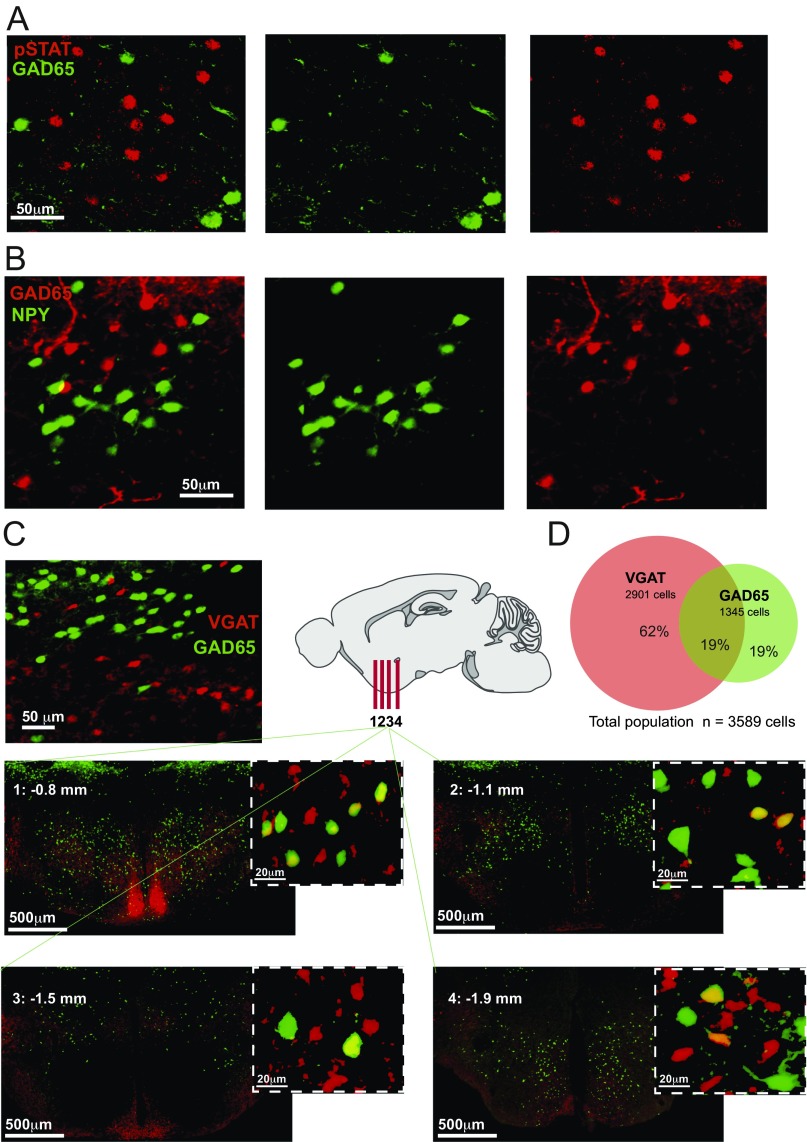
Fig. S2.
Neurochemical characteristics of GAD65LH cells. (A) Colocalization of GAD65LH cells and pSTAT3 (leptin receptor) LH cells. GAD65LH cells were labeled with GFP (GAD65-GFP transgenic mouse), and pSTAT3 neurons were labeled with Alexa Fluor 555 antibodies (Materials and Methods). A total of 11.45% of pSTAT3LH neurons contained GAD65 (analysis of 1,544 pSTAT neurons from three GAD65-GFP/pSTAT3-Alexa555 brains), and 6.89% of GAD65LH cells contained pSTAT3 (analysis of 819 GAD65 neurons from three GAD65-GFP/pSTAT3-Alexa555 brains). The % colocalization values are averages per hemisphere. (B) Colocalization of GAD65LH cells and NPYLH cells. NPY cells were labeled with GFP (NPY-hrGFP transgenic mouse), and GAD65-Ires-Cre cells were labeled with ChR2-mCherry. A total of 2.06% of NPYLH neurons contained GAD65 (analysis of 705 NPYLH neurons from three NPY-hrGFP/GAD65-mCherry brains), and 0.78% of GAD65LH cells contained NPY (analysis of 1,645 GAD65LH neurons from three NPY-hrGFP/GAD65-mCherry brains). The % colocalization values are averages per hemisphere. (C) Colocalization of GAD65LH cells and VGATLH cells. GAD65LH cells were labeled with GFP (GAD65-GFP transgenic mouse), and VGAT-Ires-Cre cells were labeled with tdTomato (CAG-tdTomato;VGAT-Ires-Cre transgenic mouse) or ChR2-mCherry (Materials and Methods). (Left) Example of LH colocalization. (Right and Bottom) More examples in coronal slices from different anteroposterior LH locations (bregma coordinates indicated on the slides). (D) Quantification of data in A (combined cell counts from three brains).

