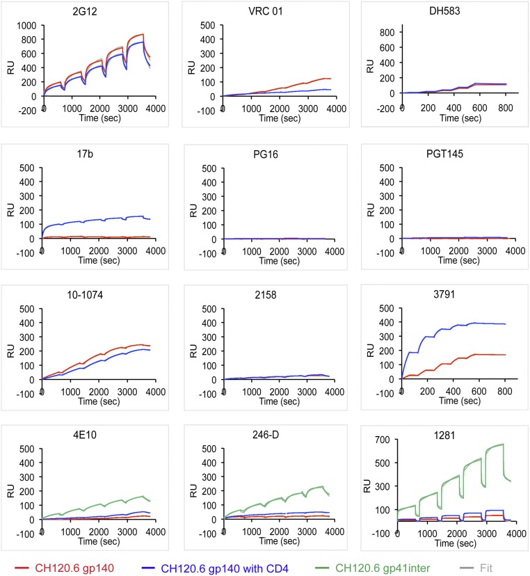
Fig. 2.
Interactions of the CH120.6 gp140 with monoclonal antibodies. Envelope proteins or their purified complexes with two-domain CD4 were captured on the surface of a sensor chip coated with an anti-histidine antibody to avoid potential artifacts introduced by protein immobilization. Fab fragments of each antibody at various concentrations were passed over the envelope trimer surface individually without regeneration for single-cycle kinetic (SCK) analysis. The recorded sensorgram for gp140 is in red, for the gp140-CD4 complex in blue, and for gp41-inter in green; the fitted curves are in gray. Sensorgrams were fit using a 1:1 binding model; binding constructs are summarized in Tables S3 and S4.

