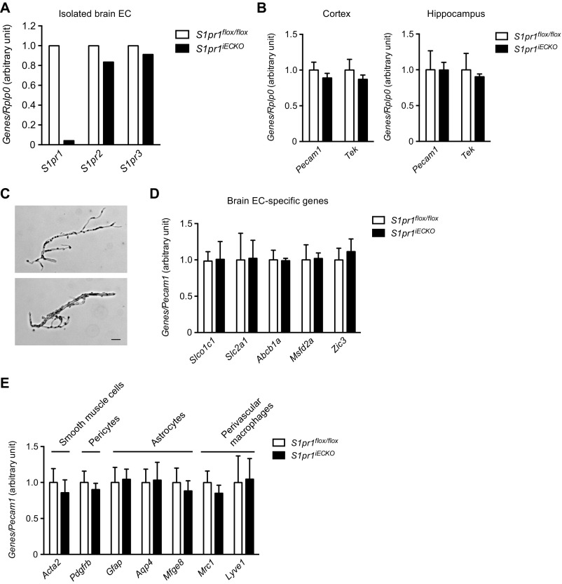
Fig. S1.
Gene expression analysis for S1P receptors, pan-EC, brain EC, and cells of the NVU. (A) qPCR analysis on the mRNA expression for major endothelial S1P receptors, S1pr1, S1pr2, and S1pr3 in isolated brain ECs from control (S1pr1flox/flox) and S1pr1iECKO mice (n = 1). (B) qPCR analysis on the mRNA expression for pan-EC maker genes, Pecam1 and Tek in cerebral cortex and hippocampus from control and S1pr1iECKO mice (n = 5). (C) Brightfield images of representative microvessels isolated from whole brain. Microvessels ranged from 4 to 15 μm in diameter. (Scale bar: 20 μm.) (D and E) qPCR analysis on the mRNA expression of genes specific for brain-EC (D), and other cell types constituting NVU; smooth muscle cells, pericytes, astrocytes, and perivascular macrophages (E) in brain microvascular fragments from control and S1pr1iECKO mice (n = 4).

