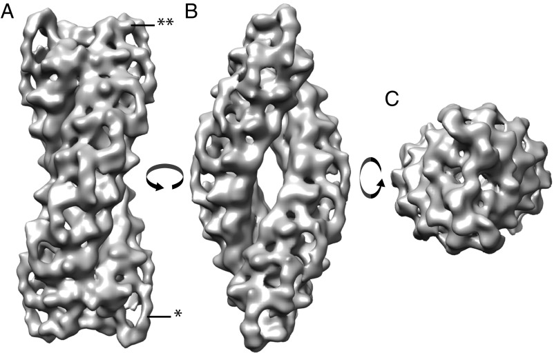
Fig. 2.
In situ structure of TPPII. Structure of the TPPII complex after subtomogram averaging. Isosurface representation of the in situ TPPII structure with B-factor applied is shown in three different views: (A) “dumbbell view,” (B) “navette view,” and (C) “top view.” The obtained TPPII structure is double-stranded and spindle-shaped and shows nine handles on each side of the strands (single asterisk). Small protrusions are resolved, which corresponds to an insertion of the catalytic Asp and His residues (DH insert highlighted by the double asterisk).

