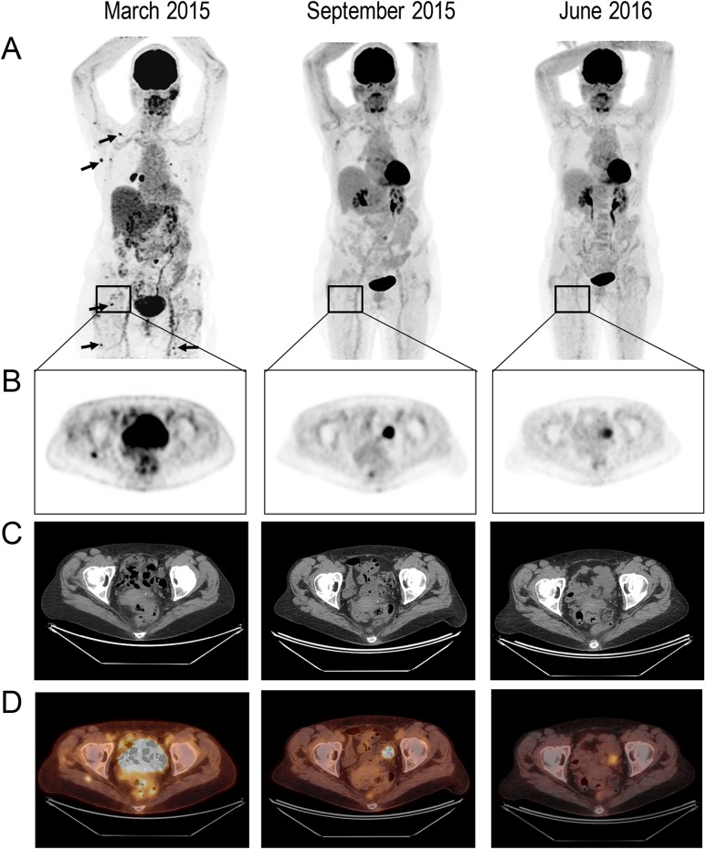
Figure 1:
Response to treatment in a breast cancer patient with diffuse skeletal muscle metastases.(A) 3D MIP (maximum intensity projection) 18F-FDG-PET (anterior views) performed before treatment (March 2015), 4 months after treatment initiation (September 2015) and 1 year on treatment (June 2016). Pretreatment images show multifocal hypermetabolic foci (arrows), from which the most intense and prominent are one in the external part of the right pectoralis major muscle and one in the right lower area of the periscapular; (B) Selected transaxial slice of a right gluteal muscle mass with increased FDG uptake; (C) the selected region slice on CT and (D) fused PET/CT.

