Figure 6b:

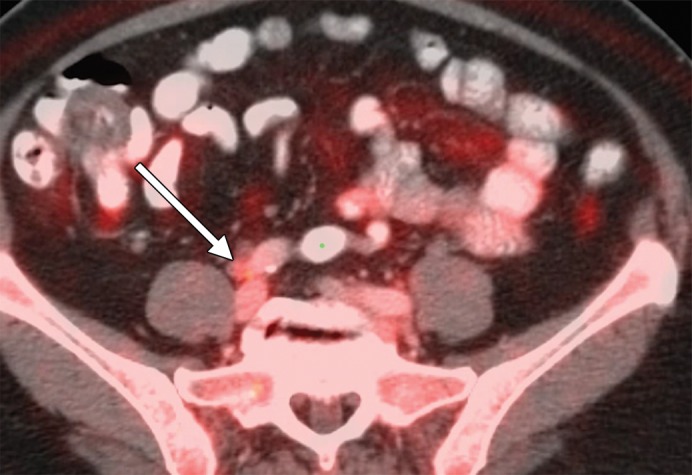
An example of a false-positive PET/CT. (a) Axial contrast-enhanced CT scan. A rounded right common iliac LN is present (arrow). (b) Axial fused PET/CT image. The LN is FDG PET avid (arrow). Pathologic findings were negative.

An example of a false-positive PET/CT. (a) Axial contrast-enhanced CT scan. A rounded right common iliac LN is present (arrow). (b) Axial fused PET/CT image. The LN is FDG PET avid (arrow). Pathologic findings were negative.