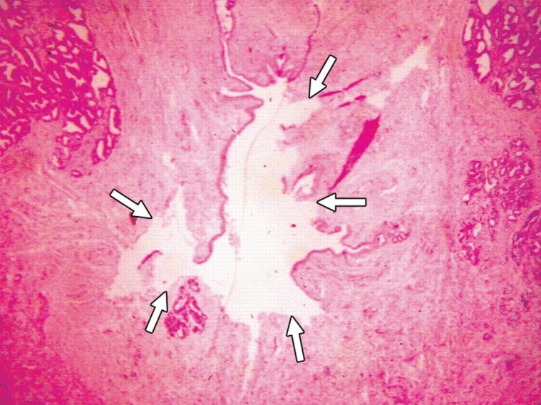
Figure 3c:

(a) Transverse contrast-enhanced pulse-inversion harmonic US image after RF ablation in a dog without any cooling protection shows no contrast enhancement of the urethra (arrowheads) and NVB (arrows). (b) Gross specimen shows thermal damage of the urethral wall (arrowheads) and NVB areas (arrows). (c) Histologic slide shows partial damage to the urethral wall (arrows). (Hematoxylin-eosin stain; original magnification, ×10.)
