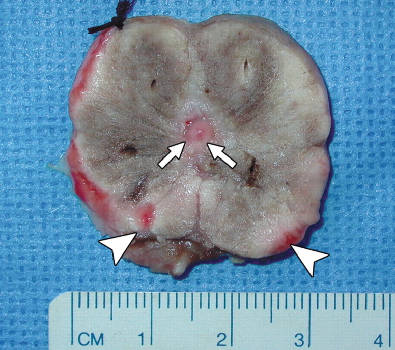
Figure 4b:

(a) Transverse contrast-enhanced pulse-inversion harmonic US after RF ablation demonstrates normal flow in the urethral wall (arrow) and the NVB areas (arrowheads) on both sides. (b) Pathologic specimen demonstrates preservation of the urethral wall (arrows) and the NVB areas (arrowheads) on both sides. Histologic slides show normal appearance of the (c) urethral wall and (d) normal NVB region (N = nerve cell, V = vessel). (Hematoxylin-eosin stain; magnification in c, ×10; magnification in d, ×40).
