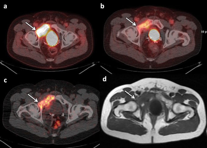
Figure 11.
FDG PET/CT, FDHT PET/CT, 89Zr-J591 PET/CT, and MR imaging of a 47-year-old man undergoing follow-up 2 years after prostatectomy and radiation therapy for prostate cancer (Gleason score, 4 + 5 = 9). Axial fused FDG PET/CT image (a), axial fused FDHT PET/CT image (b), axial fused 89Zr-J591 PET/CT image (c), and axial T1-weighted MR image (d) of the pelvis show a right superior pubic ramus metastasis extending into the acetabulum (arrow). Tracer uptake was depicted with all three PET tracers.

