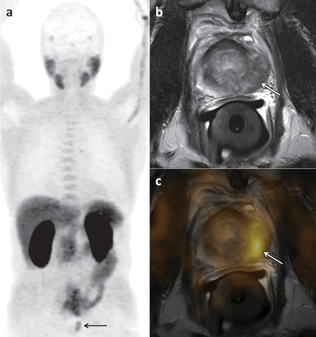
Figure 3.
18F-Choline PET/CT and MR imaging of a 66-year-old man with newly diagnosed prostate cancer and a PSA level of 30 ng/mL. (a) Early coronal maximum intensity projection image from 18F-choline PET shows unspecific focal 18F-choline uptake (arrow) in the left part of the prostate. (b) Axial T2-weighted MR image shows a hypointense lesion (arrow) in the left prostatic peripheral zone consistent with prostate cancer, a finding that was confirmed by the results of histopathologic examination. (c) Manually fused image combining axial PET image and axial MR image of the prostate depicts the 18F-choline activity (arrow) in the left peripheral zone.

