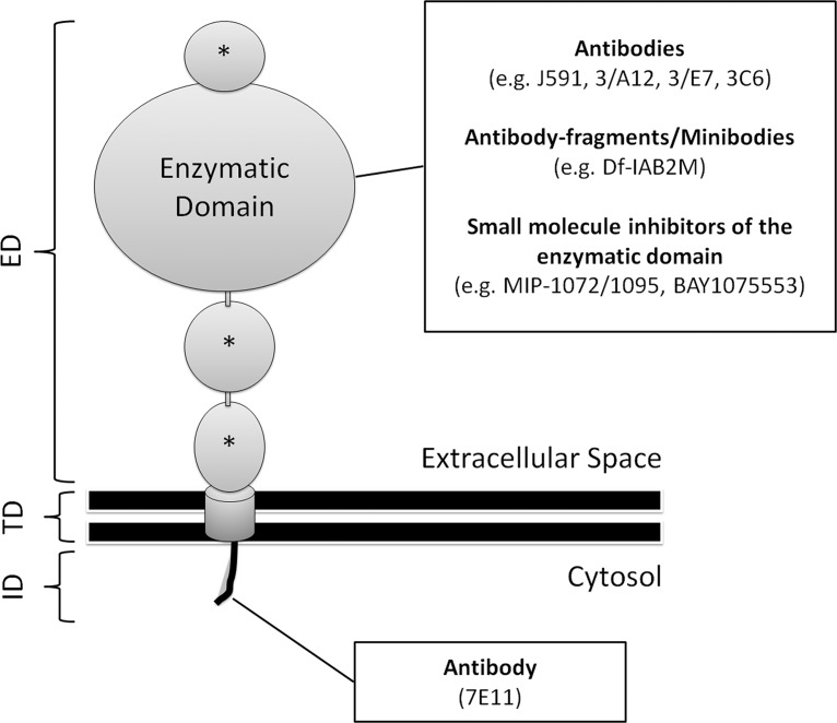
Figure 9.
Diagram of a PSMA molecule. The molecule is composed of a short intracellular domain (ID), a hydrophobic transmembranous domain (TD), and a large extracellular domain (ED). The latter consists of a large enzymatic portion and three smaller domains (*), the functions of which are not known. PSMA-directed imaging tracers can be divided into those targeting the ID and those that bind to the ED or inhibit its enzymatic domain. The ID contains a motif that is responsible for the internalization of the molecule into the endosomal recycling system of the cell.

