
Figure 2. EZH2 is predominantly expressed in neurons in the spinal dorsal horn under normal conditions, and nerve injury increases the number of neurons with EZH2 expression and microglia with EZH2 expression.
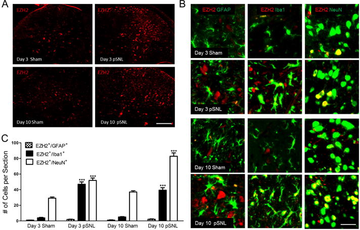
(A) shows samples of EZH2 staining in L4–L5 spinal dorsal horn ipsilateral to the operation site in sham and pSNL rats on days 3 and 10 after surgery. Scale bar: 200 μm. (B) shows co-localization of EZH2 staining (red) with different cellular markers (in green): Iba1 for microglia, GFAP for astrocytes, and NeuN for neurons. Scale bar: 50 μm. (C) Bar graphs show the mean (+ S.E.) numbers of EZH2 +/Iba1+, EZH2+/GFAP+, and EZH2+/NeuN+ cells in the spinal dorsal horn per section in sham operated and pSNL rats on days 3 and 10 after surgery. Four to five rats per group were used for analysis. Comparisons of the same cellular types on the same days after surgery between sham-operated and pSNL rats are shown. *** p < 0.001.