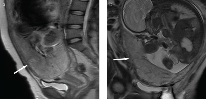
Figure 3. a, b.
A 20-year-old woman with complete placenta previa. Sagittal (a) and coronal (b) T2-weighted images showing the focally interrupted placenta/myometrial interface (arrow) in the lower anterior segment of the uterus without appearance of low signal intensity bands on T2-weighted imaging. The patient had minor hemorrhage of 800 mL, and thus uterus was preserved.

