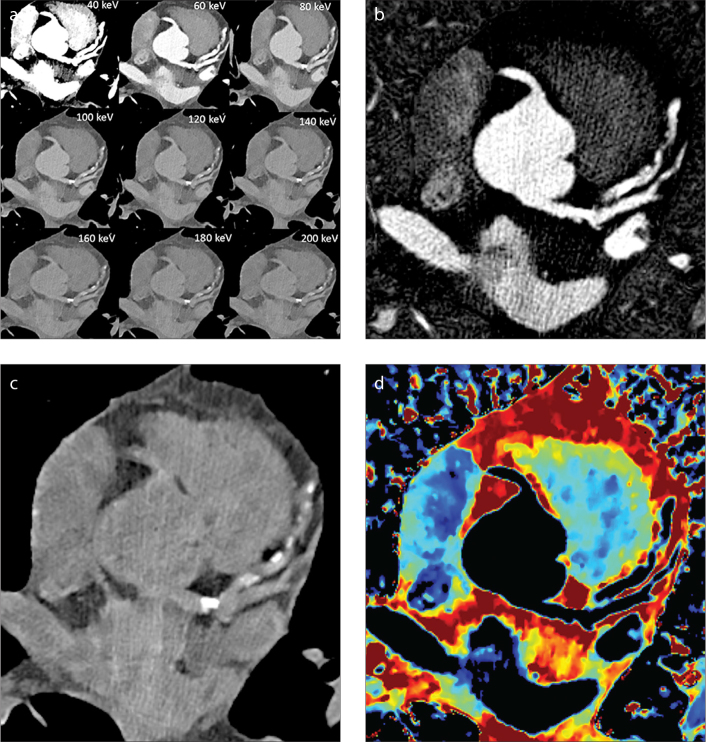
Figure 2. a–d.
Types of spectral images. Panel (a) shows cross-section of the heart displayed as virtual monoenergetic images from 40 to 200 keV displaying tissue attenuation properties similar to those resulting from imaging with a monoenergetic beam at a single keV level. Panel (b) shows iodine density maps in which pixels containing iodine are preserved but all other pixels appear black. Panel (c) displays virtual unenhanced images in which pixels containing iodine have been removed. Panel (d) shows effective atomic number (Zeffective) images; pixel values equal Zeffective of tissue contained within each voxel.

